PET / CT OVERVIEW
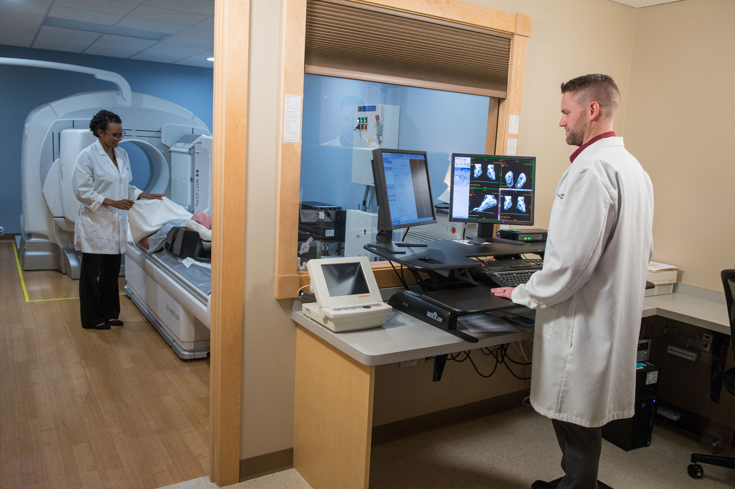
Positron Emission Tomography (PET)/Computed Tomography (CT) technology is an advanced nuclear imaging exam that allows physicians to view and measure not only the size, shape, and location of an organ or internal structure but also its function, in one scan.
Computed Tomography (CT) uses x-ray technology and computers to view the anatomy of tissues with exceptional clarity and detail. Positron Emission Tomography (PET) captures images of the biological function of tissues by detecting the distribution of a radioactive substance that is injected into the patient prior to the exam.
PET/CT scans are a valuable tool in the detection and diagnosis of cancer, blocked arteries in the heart and neurological conditions such as Alzheimer’s and dementia.
What should I expect?
Typically, PET/CT scans begin with an injection of a radioactive sugar solution. Following the injection, the patient rests in a private room for approximately 60 minutes.
As your body metabolizes the sugar, the positrons that are attached to the sugar can be seen by the PET/CT scanner. Areas with higher radioactivity will show up as “hot spots” on a PET/CT image, which can be an indication of rapidly growing tumors as cancerous cells generally consume more sugar than normal tissue.
Generally, it is safe to assume up to two hours to complete the entire exam.
The PET exam itself causes no side effects and patients are able to drive themselves home if they did not require/request any sedation. If patients do opt for a mild sedative to relieve anxiety etc., they are required to make transportation arrangements for a ride after the exam.